Please join us for a Frontiers in Microscopy online talk. All welcome!
Date/Time: Wednesday, 12th February 2025, 1:30pm – 2:30pm (AEST)
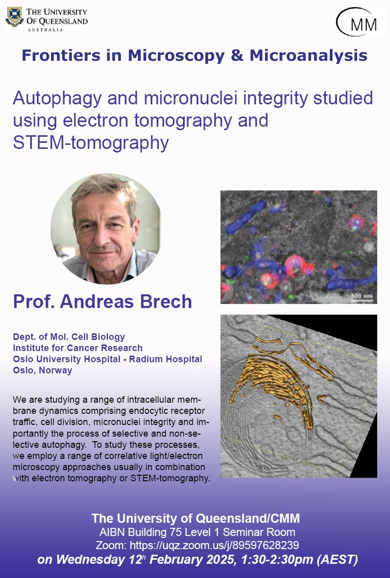
Title: Autophagy and micronuclei integrity studied using electron tomography and STEM-tomography
Presenter: Prof. Andreas Brech, Advanced Electron Microscopy Core Facility, Oslo University Hospital
Online: https://uqz.zoom.us/j/89597628239
We are studying a range of intracellular membrane dynamics comprising endocytic receptor traffic, cell division, micronuclei integrity and importantly the process of selective and non-selective autophagy. The autophagic process is controlled by several well-characterized proteins (ATG-proteins) but is also governed by the physical properties of the lipid membranes involved. We have shown that liquid condensates can be turned over by a process termed piecemeal autophagy, which is strongly determined by lipid membrane properties (in collaboration with R. Fromm). The autophagic adaptor protein p62 is one of the key players in this system and we are currently trying to understand how this condensation-prone protein undergoes phase separation and interplays with autophagic dynamics. We are also speculating whether phase separation could be an important mechanism during micronuclei rupture and that an ESCRT-driven repair process is hampered due to nuclear membrane behavior in relation to possible condensation. To study these processes, we employ a range of correlative light/electron microscopy approaches usually in combination with electron tomography or STEM-tomography. We have recently explored possibilities to acquire tomograms of large cellular volumes to further characterize autophagy by STEM-tomography. The great advantage of STEM-tomography over regular electron tomography is that much thicker samples (up to 1500 nm) can be observed increasing the imaged volume in each tilt series by a factor of 4. This can be performed with a pixels size down to 1 nm, thereby filling a niche in volume EM which is currently not reachable with FIB-SEM due to the limitations in resolution (> 4 nm pixel size).