On Wednesday the 8th of February, we will be joined for a Frontiers in Microscopy seminar by Dr Yannick Schwab, Team Leader and Head of Electron Microscopy Core Facility at EMBL Heidelberg. He will be presenting some recent research revolving around precise targeting for volume electron microscopy.
Date/Time: Wednesday 8th February, 14:00 to 15:00 (AEST)
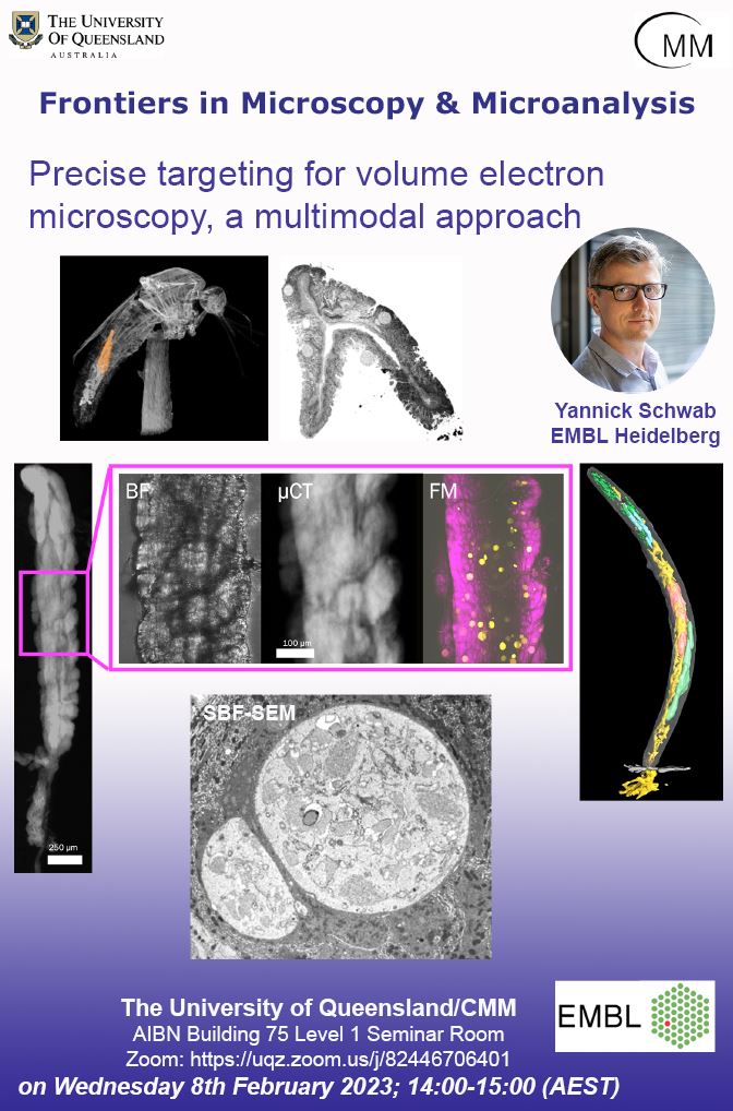
Title: Precise targeting for volume electron microscopy, a multimodal approach
Presenter: Dr Yannick Schwab, EMBL Heidelberg
Venue: AIBN Building 75, Seminar Room Level 1
Zoom: https://uqz.zoom.us/j/82446706401
Volume electron microscopy methods (vEM) are powerful to study the complex ultrastructure of cells. Based on serial imaging of either resin sections (ssTEM, ssET and array tomography) or of sequentially ablated bock surface (SBEM and FIB-SEM), they uniquely capture cells and organelles shapes and interactions in three dimensions. Resulting datasets can lead to detailed morphological quantifications. High resolution vEM are often performed on large samples, whether multicellular organisms or tissues, which opens to studying cell-cell interactions within their micro-environment. In some cases, such analyses are performed on a subset of cells, selected for their particular phenotype or for their identity. Targeting the acquisition to these regions is thus interesting as it optimizes dramatically the acquisition time, the sampling throughput and the amount of data generated. Because direct targeting within the embedded specimen is almost impossible at the EM, multimodal correlative methods have been developed to establish with precision the position of the volume of interest.
This talk will describe the targeting methods that are developed at EMBL. They rely on 3D maps built from fluorescence microscopy or X-ray imaging, and on specific workflows to accurately and semi-automatically approach the regions of interest prior to EM imaging. Example applications will show how to image selected regions of interest in multiple specimens including models used to study different infection stages of the malaria parasite and plankton cells collected in the field.