We would like to invite you to our first 2020 Frontiers in Microscopy & Microanalysis talk this Friday starting at 1pm.
Title: Cryo Micro Electron Diffraction of Proteins, by two guests – Dr Mathieu Coinçon & Dr Max Clabbers from Stockholm University
Date/time: Friday 7th February, 1pm – 2pm
Venue: AIBN (Building #75) Level 1 Seminar Room
Flyer
Abstracts:
1pm to 1.30pm
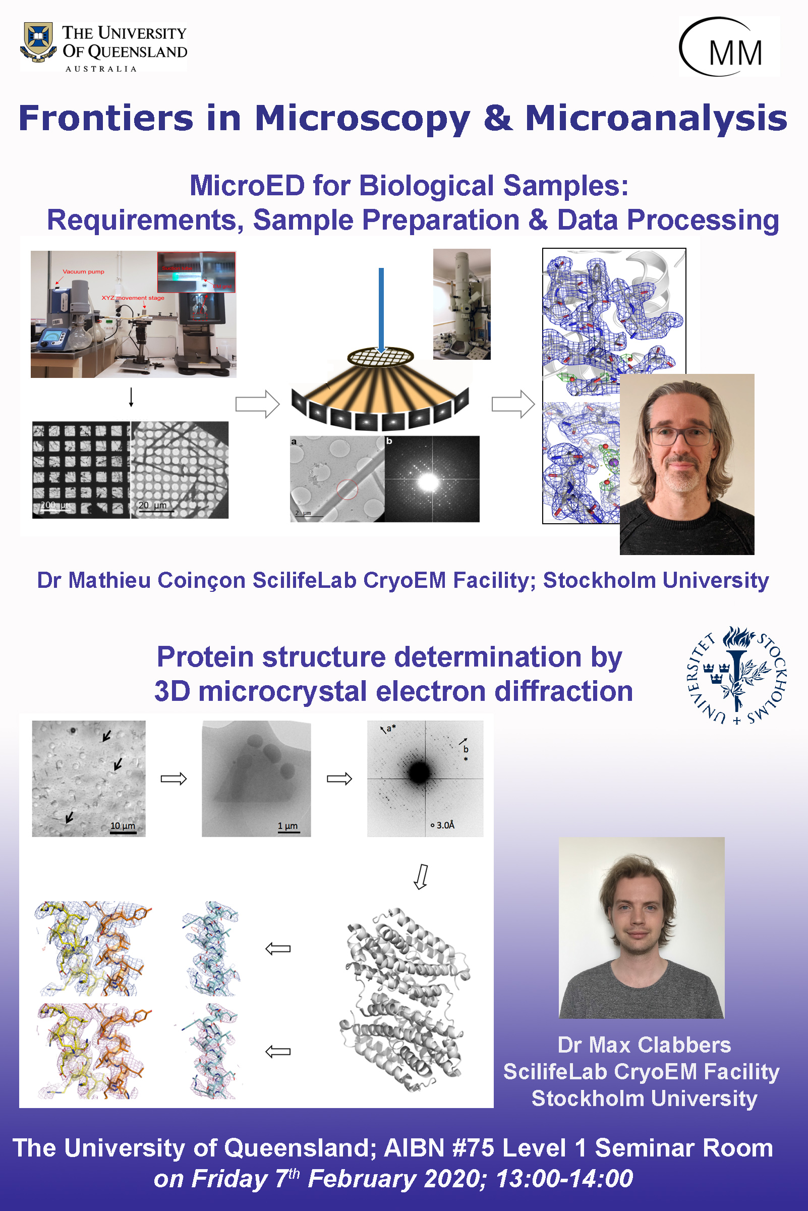
MicroED for Biological Samples: Requirements, Sample Preparation and Data Processing
Dr. Mathieu Coinçon; ScilifeLab CryoEM Facility; Stockholm University
3D electron diffraction techniques used for decades in material science were recently adapted to study biological samples. Microcrystal electron diffraction (MicroED) enables crystals considered too small (sub-microns) for conventional X-ray diffraction to be used to solve protein structures1-7.
We will present how to:
- choose / grow crystals suitable for electron diffraction
- prepare the sample grids
- diffract these crystals under the electron beam using the (continuous) rotation method2,3
- process the data using standard crystallographic routines8
We will focus especially on techniques and software developed in the lab.
- InsteaDMatic9: A DigitalMicrograph script developed to coordinate TEM goniometer rotation and detector image recording for continuous rotational electron diffraction (cRED) data acquisition.
- Preassis10: a simple and newly developed pressure-assisted method for preparing cryo-EM specimens of both single particles and micro-crystals. By using Preassis, suitable cryo-EM specimens can be prepared from proteins with concentrations as low as 0.18 mg/ml. In addition, Preassis can handle protein crystal suspensions with both low and high viscosity. More importantly, the method is simple and easy to implement, and no special EM grids are required. With minor modifications, the Preassis setup can be adapted to existing cryo-EM vitrification devices, making it widely accessible to various cryo-EM labs.
1.30pm to 2pm
Protein structure determination by 3D microcrystal electron diffraction
Dr. Max Clabbers, ScilifeLab CryoEM Facility; Stockholm University
Microcrystal electron diffraction (MicroED) has recently shown potential for structural biology, enabling macromolecular structure determination when only (sub-) micron sized 3D crystals are available that are beyond what can be resolved by conventional x-ray crystallography1–7. MicroED data can efficiently be collected using the (continuous) rotation method2,3, and standard crystallographic routines for data processing can be adapted8. The sample preparation is similar to that of cryo-EM, making MicroED highly adaptable and complementary to existing techniques. However, up until now MicroED had only been applied to refine already known protein structures that were previously solved by x-ray diffraction. Here, we present a first unknown protein structure of a SaR2lox metelloenzyne solved using MicroED9. Despite encountering preferential orientation of the plate-like crystals, low signal-to-noise ratio, and a highly viscous sample environment, the structure could successfully be solved by molecular replacement using a search model of only 35% sequence identity. The resulting electrostatic scattering potential map at 3.0Å resolution was of sufficient quality to allow accurate model building and refinement. Furthermore, we present the structure of the Toll/interleukin-1 receptor (TIR) domain of the signalling adaptor protein MyD88. We used MicroED to solve the structure of MyD88TIR from thin filaments of typically 50-150 nm diameter10. The resulting map enabled accurate modelling of the structure using reciprocal space methods and rebuilding via interactive molecular dynamics11. Several loop regions in our MicroED model were remodelled compared to previously reported structures12,13, providing novel insights into the structural basis of signalling via TIR domain binding interactions. Our results illustrate that MicroED has the potential to become a widely applicable technique for structural biology, complementing x-ray crystallography when crystal volume is the limiting factor inhibiting structure elucidation.
All are welcome!


