Please join us for a Frontiers in Microscopy Seminar on Monday, 20 November.
Date/Time: Monday, 20 November 2023, 15:30 – 16:30 (AEST)
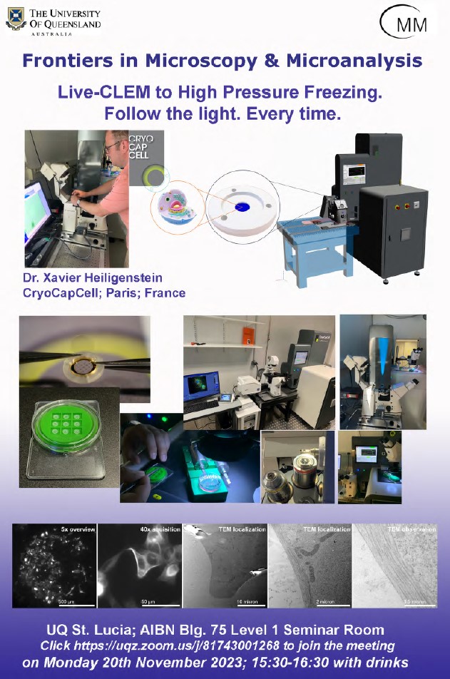
Title: Live-CLEM to High Pressure Freezing. Follow the light. Every time.
Presenter: Dr Xavier Heiligenstein, CryoCapCell, Paris, France
Venue: AIBN Building 75, Level 1 Seminar Room
Zoom: https://uqz.zoom.us/j/81743001268
Abstract:
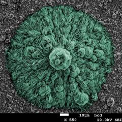
Correlative light and electron microscopy (CLEM) is an advanced imaging technique that combines the strengths of light microscopy (LM) and electron microscopy (EM) to provide a comprehensive understanding of biological structures and processes. To preserve samples for EM, high pressure freezing (HPF) is utilized, which rapidly freezes them under high pressure to minimize damage caused by ice crystal formation.
To achieve Live-CLEM HPF, with optimal sample preservation, we have developed a unique pipeline, from sample culture (using the CryoCapsule) and live observation (LSM900 Zeiss Airyscan), to HPF vitrification (HPM Live µ) in less than 2 seconds. Subsequent preparation for electron microscopy (including freeze substitution in FS basket and embedding in electro-conductive resin R221) reveal fine structural details of any of the live-imaged sample.
The high reliability of our pipeline guaranties us to retrieve 100% of the live-tracked cell in the TEM, with optimal preservation. This opens a novel frontier for dynamic CLEM.
All welcome!
Some background information on the speaker:
Xavier Heiligenstein, PhD
During my PhD and following years, I invented the CryoCapsule® (Heiligenstein, Heiligenstein, et al., 2014; Heiligenstein, Hurbain, et al., 2014) and co-developed new tools for CLEM:
- The eC-CLEM software facilitates the fusion of data collected from different devices into a single image file to validate the localization of a fluorescence signal in a set of electron microscopy images (Heiligenstein, Paul-Gilloteaux, Raposo, & Salamero, 2017; Paul-Gilloteaux et al., 2017).
- In order to optimize the use of the CryoCapsule and improve the spatio-temporal accuracy of live imaging with electron microscopy, we invented a high-pressure freezer (cryo-immobilization of samples) combined with a photon microscope: the HPM Live µ (Heiligenstein et al., 2021).
- Finally, to embrace the development of volumetric electron microscopy (vEM) and optimize multimodal imaging protocols (optical and electronic imaging of the same sample included in resin), we created a new electro-conductive resin, R221 (Heiligenstein et al., 2021; Heiligenstein & Lucas, 2022).
- In 2013, to commercialize and diffuse these various tools, I co-founded the CryoCapCell company (www.cryocapcell.com)
- Since 2021, we have installed our research group at the INSERM U1195 Unit to continuously interact with research scientist and remain close to leading edge research with stimulating scientific questions.
In summary, during my PhD, I invented the CryoCapsule® and contributed to the development of new tools for correlative light and electron microscopy, including eC-CLEM software, a high-pressure freezer (HPM Live µ), and an electro-conductive resin (R221) for multimodal imaging. These tools enable accurate and comprehensive imaging of biological samples and provide a significant advancement in the field of microscopy.